コレクション oil red o staining protocol for cytology 121227-Oil red o staining protocol for cytology

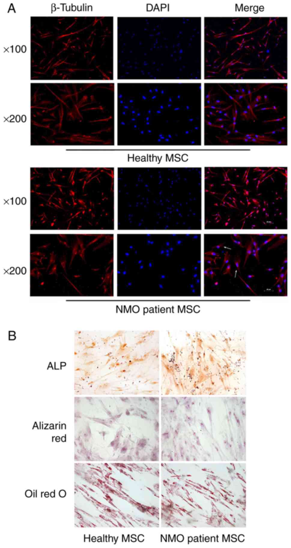
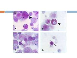
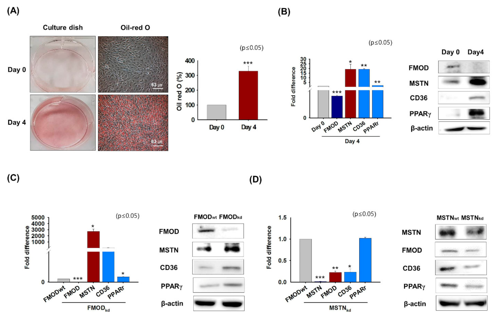
Oil Red O Staining And Morphological Changes In Dfat Gfp And 3t3 L1 Download Scientific Diagram
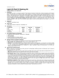
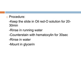
Add Oil Red O working solution for 10 min (do not touch walls of the wells) Remove all Oil Red O and IMMEDIATELY adddH2O, wash with H2O 4 times (you can wash under running tap water) Take picktures if desired Remove all water and let dry Elute Oil Red O by adding 100% isopropanol, incubate about 10 m in (can be longer)Protocol Wash Cells 2X with 1xPBS Remove media by carefully aspirating Do not touch cells (leave the chambers on!) Oil Red O Lipid Staining 10 min Oil Red O is kept as 04% Oil Red in 100% Isopropanol;
Oil red o staining protocol for cytology
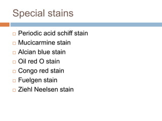
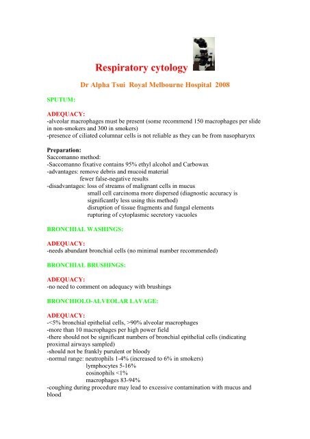
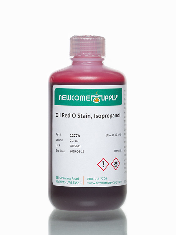
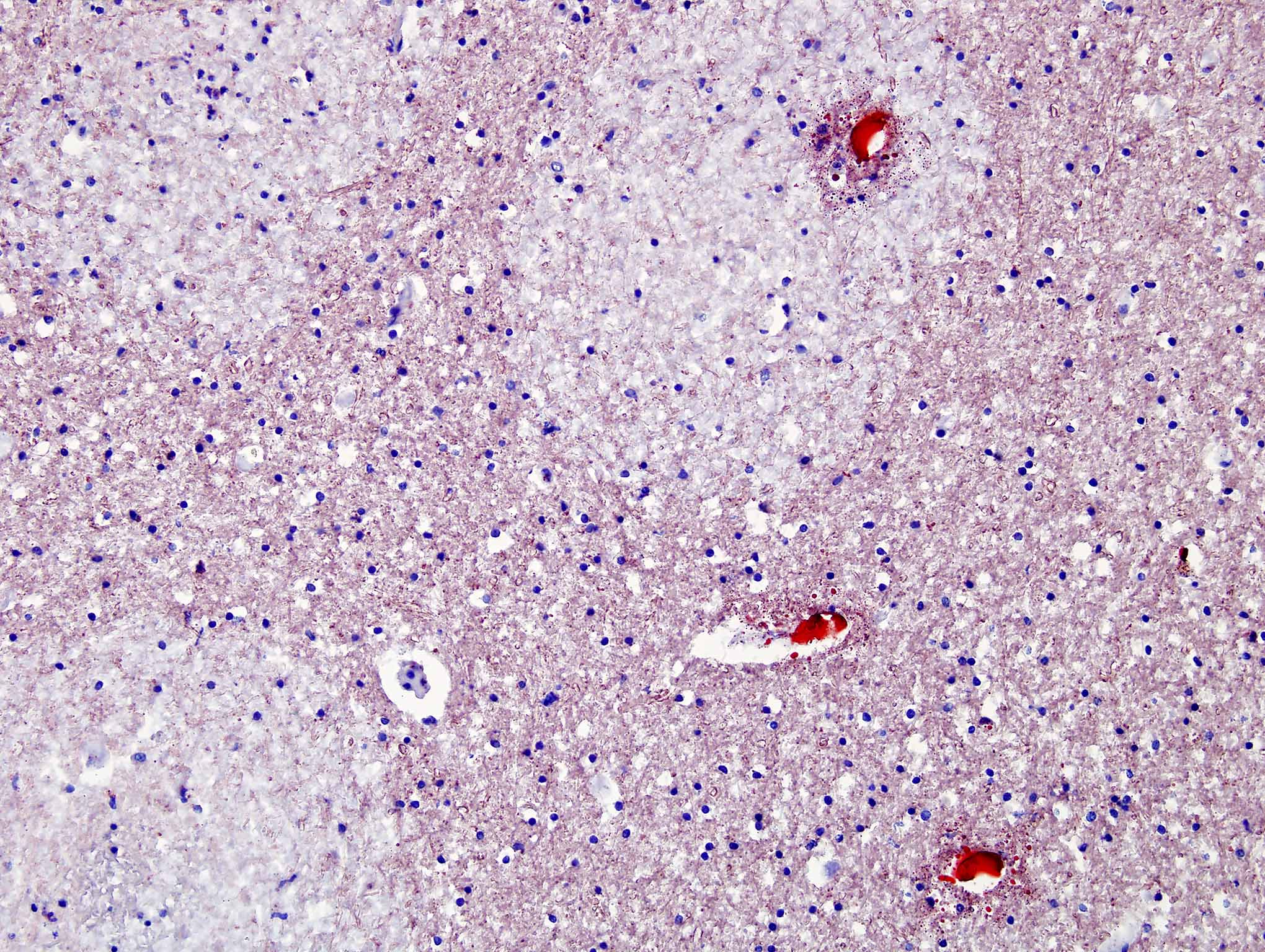
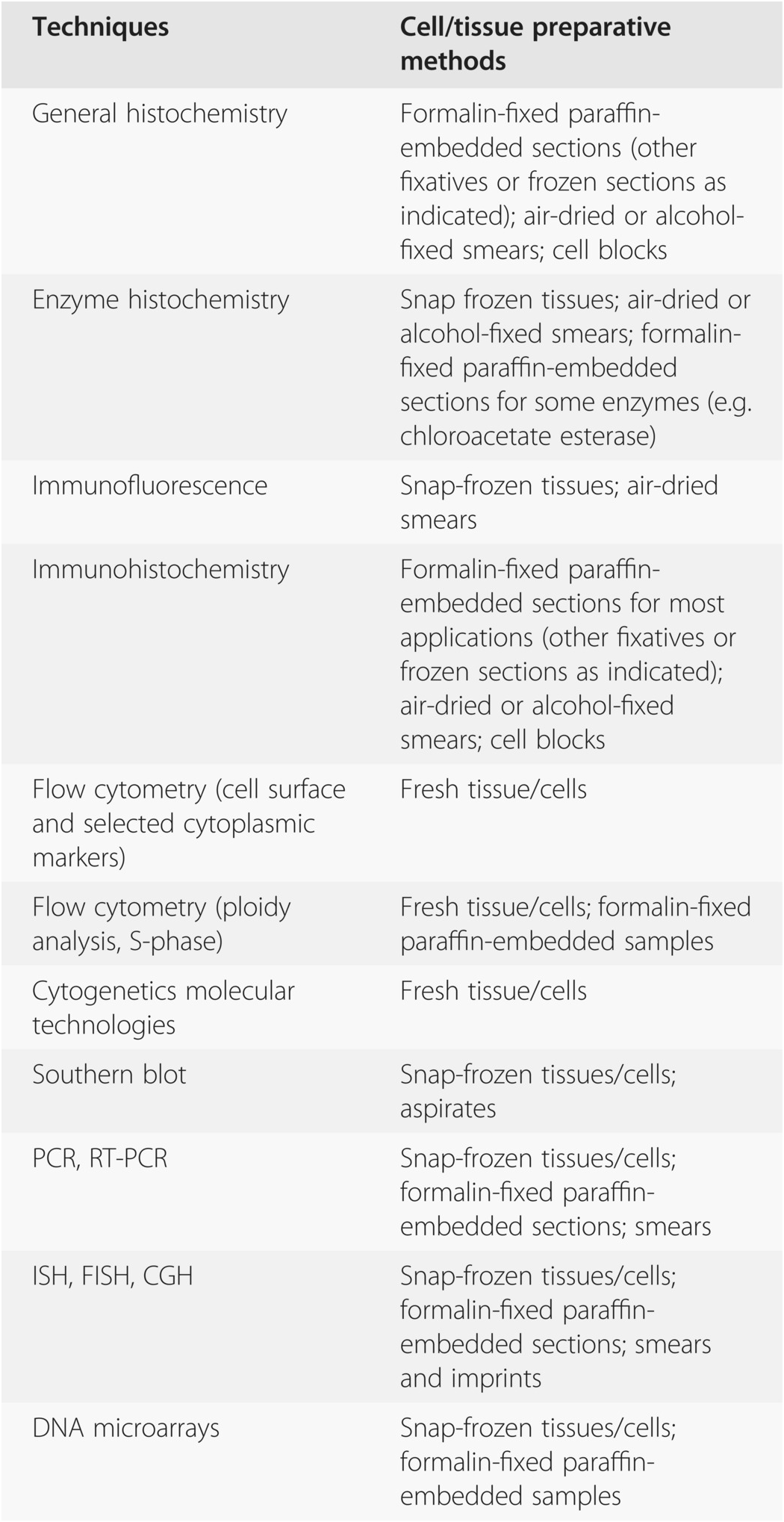
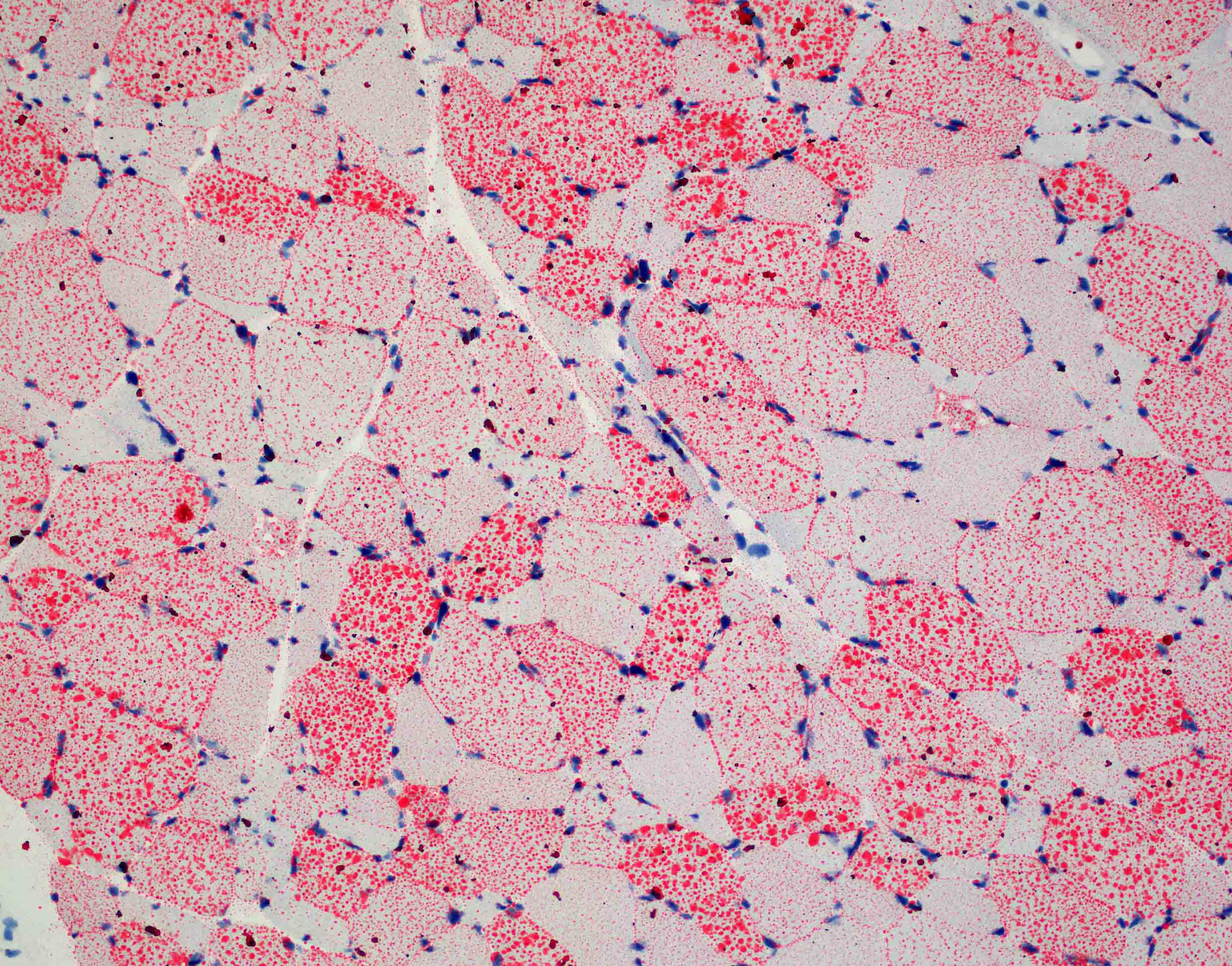
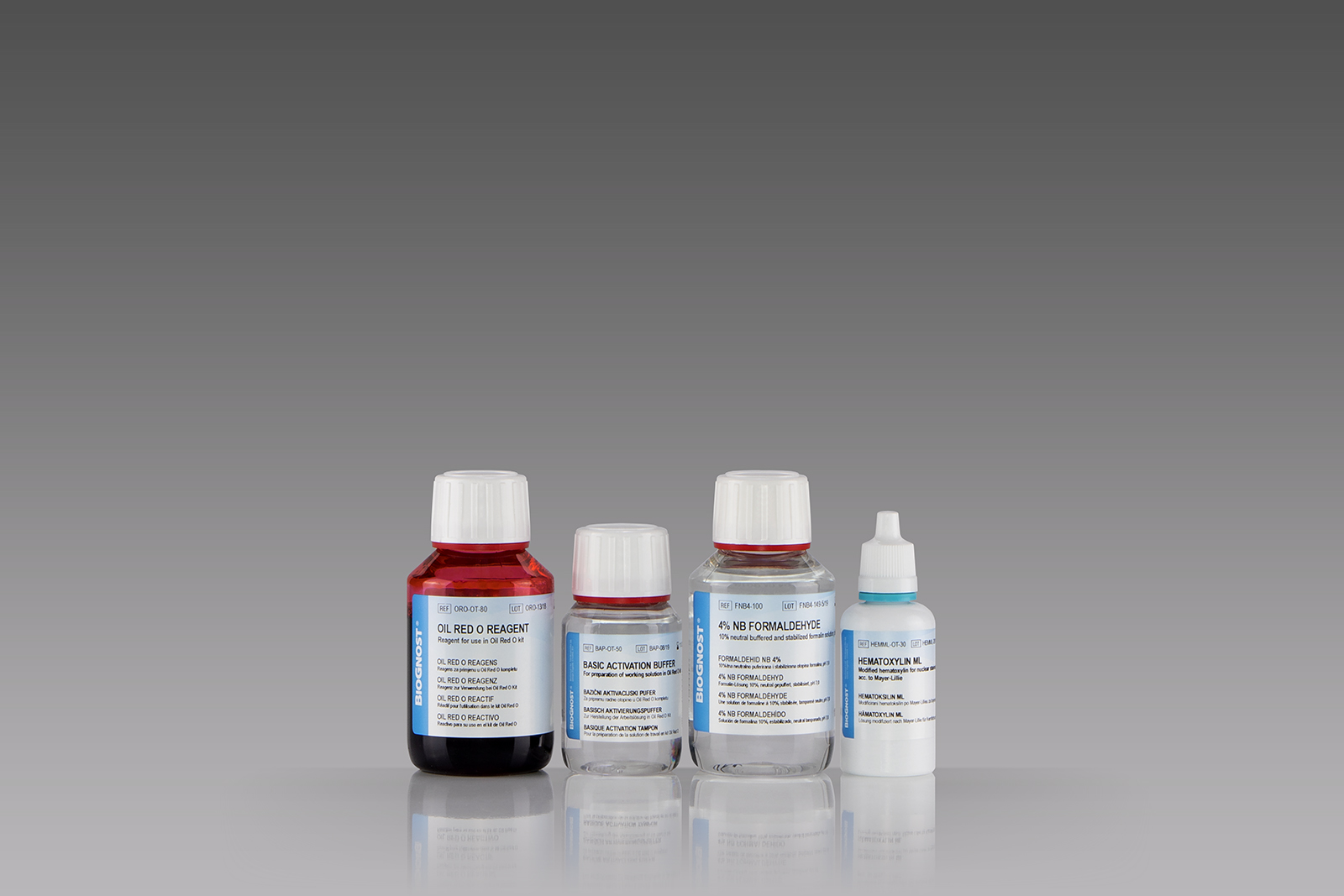
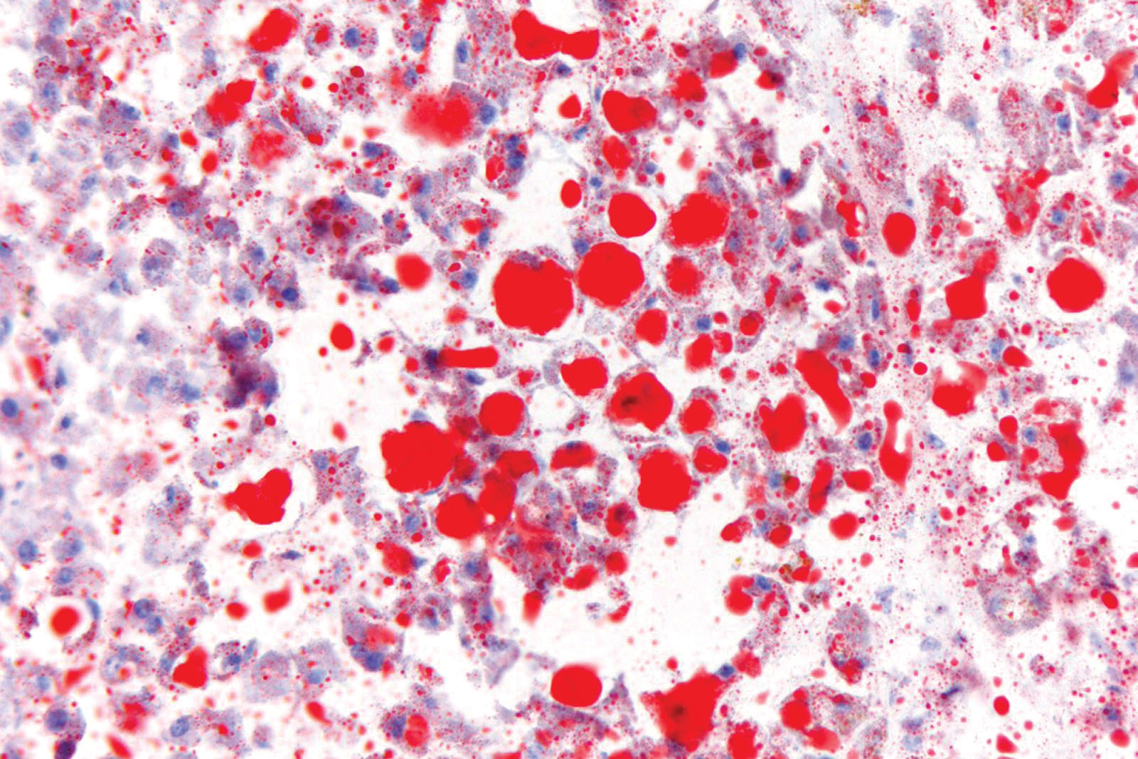
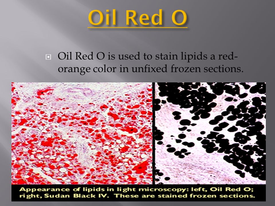
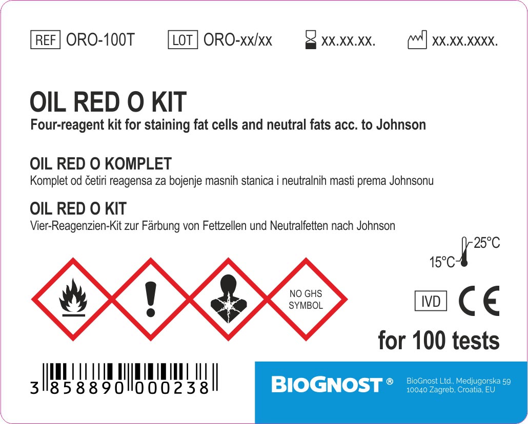
Oil red o staining protocol for cytology-1 Role of Diagnostic Cytology 09 2 Collection and Preparation 10 of Material for Cytodiagnosis 3 Cytopreparatory techniques 24 of Serous Effusions 4 Fixation of Cytology specimens 27 5 Staining methods in Cytology 30 Appendix Organisation of 36 Cytopathology Laboratory Minimum requirements for 40 setting up a small Cytology LaboratoryOil red O staining is used for the detection of neutral fat or polyethylene granula in e g histological tissues of human origin, in joint pathology or in cytological sample material The stain is carried out on cryo sections of native tissue Approx 0 stains can be performed with one 250 ml bottle This product is registered as IVD and CE
2
RamirezZacarias, JL, CastroMunozledo, F & KuriHarcuch, W Quantitation of adipose conversion and triglycerides by staining intracytoplasmic lipids with Oil red O Histochemistry 97, 493Oil red staining for semiquantitative scoring of adipogenesis Before staining, cells were washed twice with PBS and fixed in 10% (v/v) formalin (Biostain Ready Reagents, Manchester, UK) AfterMethod 1 Cut frozen sections at 8 to10mm, air dry the sections to the slides 2 Fix in formalin, briefly wash with running tap water 110 mins 3 Rinse with 60% isopropanol 4 Stain with freshly prepared Oil Red O working solution 15 mins 5 Rinse with 60% isopropanol 6 Lightly stain nuclei with alum haematoxylin 5 dips 7
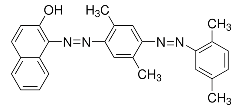
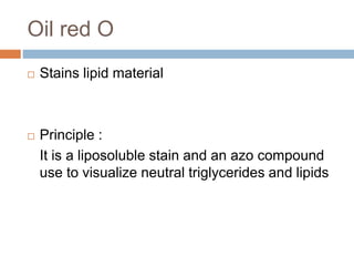
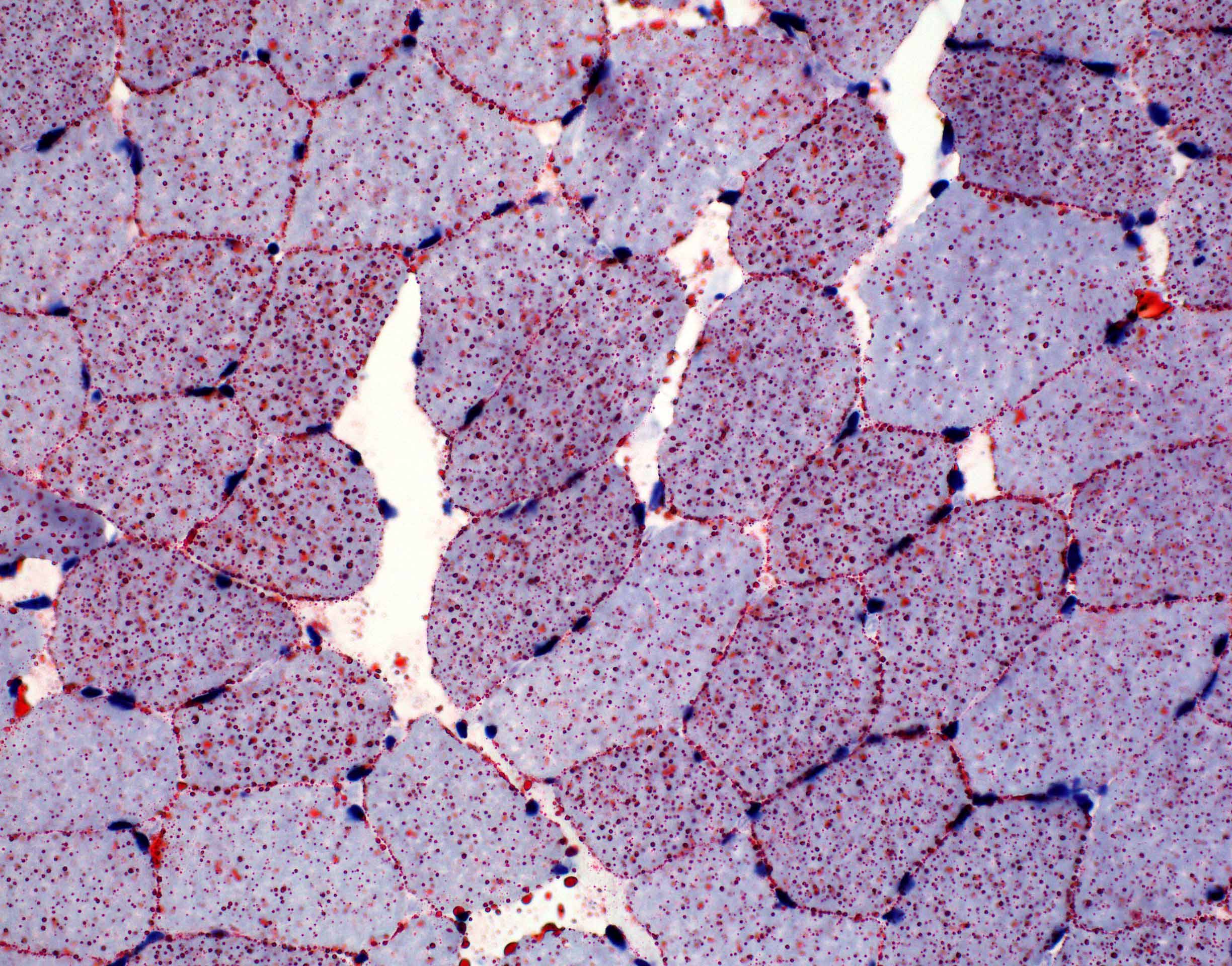
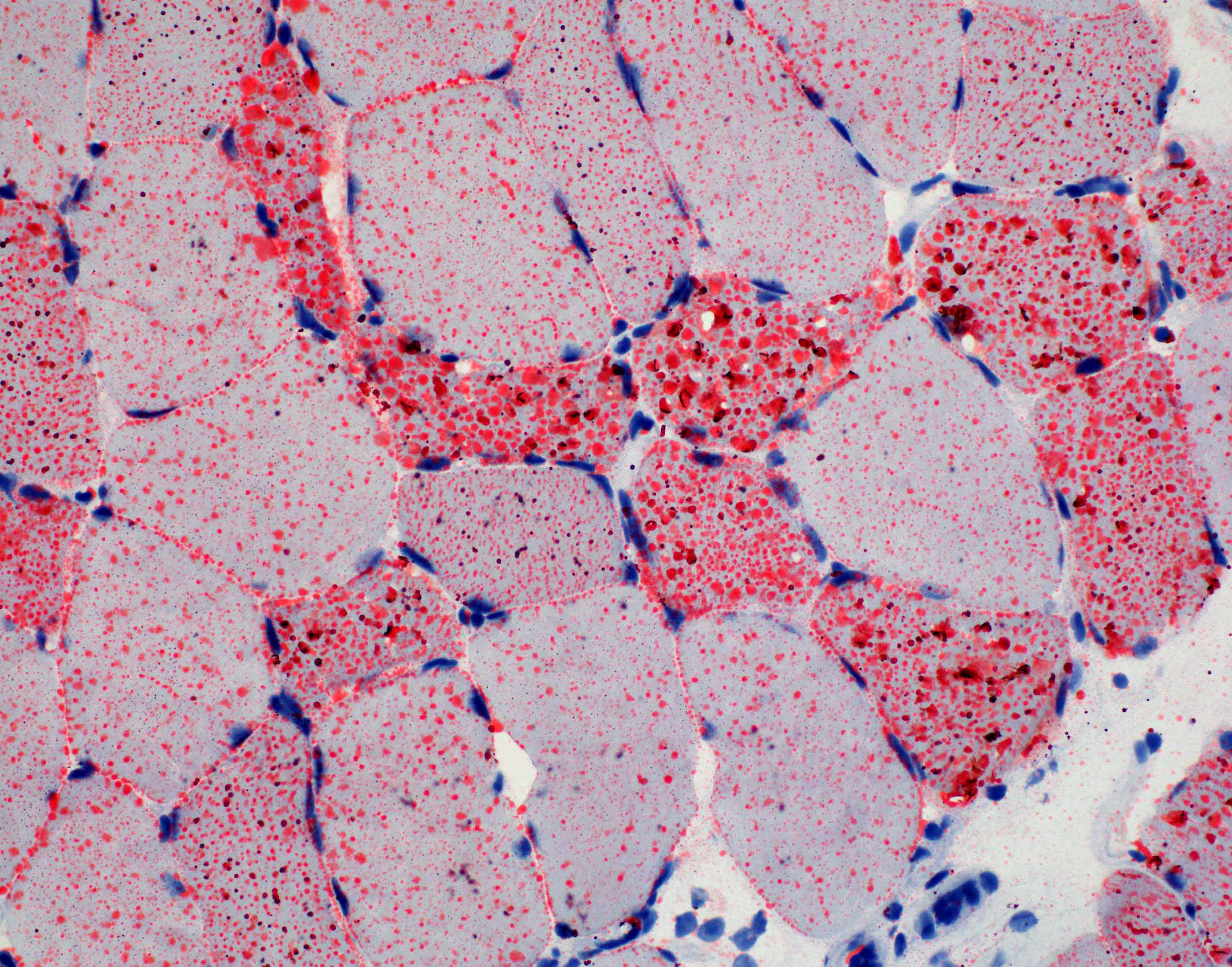
Procedure Place frozen sections at RT 30min Preheat Oil Red O Stain to 60°C Place frozen sections in 10% Formalin for 25 minutes then rinse slide in tap water (fixed slide may skip this step) Circle the tissue by Dako Pen Place slide in Propylene glycol for 2 minutes Place slide in Oil Red O Stain heated to 60°C for 6 minutes For the Oil Red O staining technique, various organic solvents, as propylene glycol or isopropyl alcohol, have been proven to successfully dissolve Oil Preparing oil red o stain Prepare the stock solution by weighing out 300 mg of oil red o powder and adding this to 100 ml of 99% isopropanol This solution is stable for one year from the date on which it is prepared In the fume hood, mix 3 parts (30 ml) of oil red o stock solution with 2 parts ( ml) deionized water and allow to sit at room
Oil red o staining protocol for cytologyのギャラリー
各画像をクリックすると、ダウンロードまたは拡大表示できます
 |  |  |
 |  |  |
 |  | |
「Oil red o staining protocol for cytology」の画像ギャラリー、詳細は各画像をクリックしてください。
 | ||
 |  |  |
 |  |  |
「Oil red o staining protocol for cytology」の画像ギャラリー、詳細は各画像をクリックしてください。
 |  |  |
 |  |  |
 |  |  |
「Oil red o staining protocol for cytology」の画像ギャラリー、詳細は各画像をクリックしてください。
 |  |  |
 |  |  |
 |  |  |
「Oil red o staining protocol for cytology」の画像ギャラリー、詳細は各画像をクリックしてください。
 |  | |
 |  | |
 |  | |
「Oil red o staining protocol for cytology」の画像ギャラリー、詳細は各画像をクリックしてください。
 |  |  |
 | ||
 |  |  |
「Oil red o staining protocol for cytology」の画像ギャラリー、詳細は各画像をクリックしてください。
 |  | |
 |  | |
 | ||
「Oil red o staining protocol for cytology」の画像ギャラリー、詳細は各画像をクリックしてください。
 |  | |
 |  | |
 |  | |
「Oil red o staining protocol for cytology」の画像ギャラリー、詳細は各画像をクリックしてください。
 |  |  |
 |  |  |
 |  |  |
「Oil red o staining protocol for cytology」の画像ギャラリー、詳細は各画像をクリックしてください。
 |  |  |
 |  | |
 |  |  |
「Oil red o staining protocol for cytology」の画像ギャラリー、詳細は各画像をクリックしてください。
 |  |  |
 |  | |
 |  |  |
「Oil red o staining protocol for cytology」の画像ギャラリー、詳細は各画像をクリックしてください。
 |  |  |
 |  |
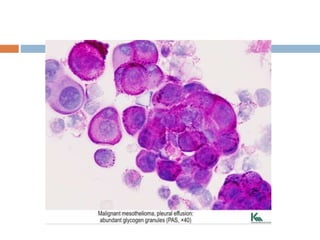
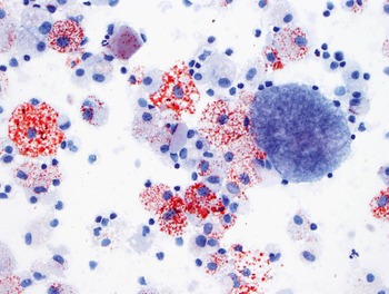
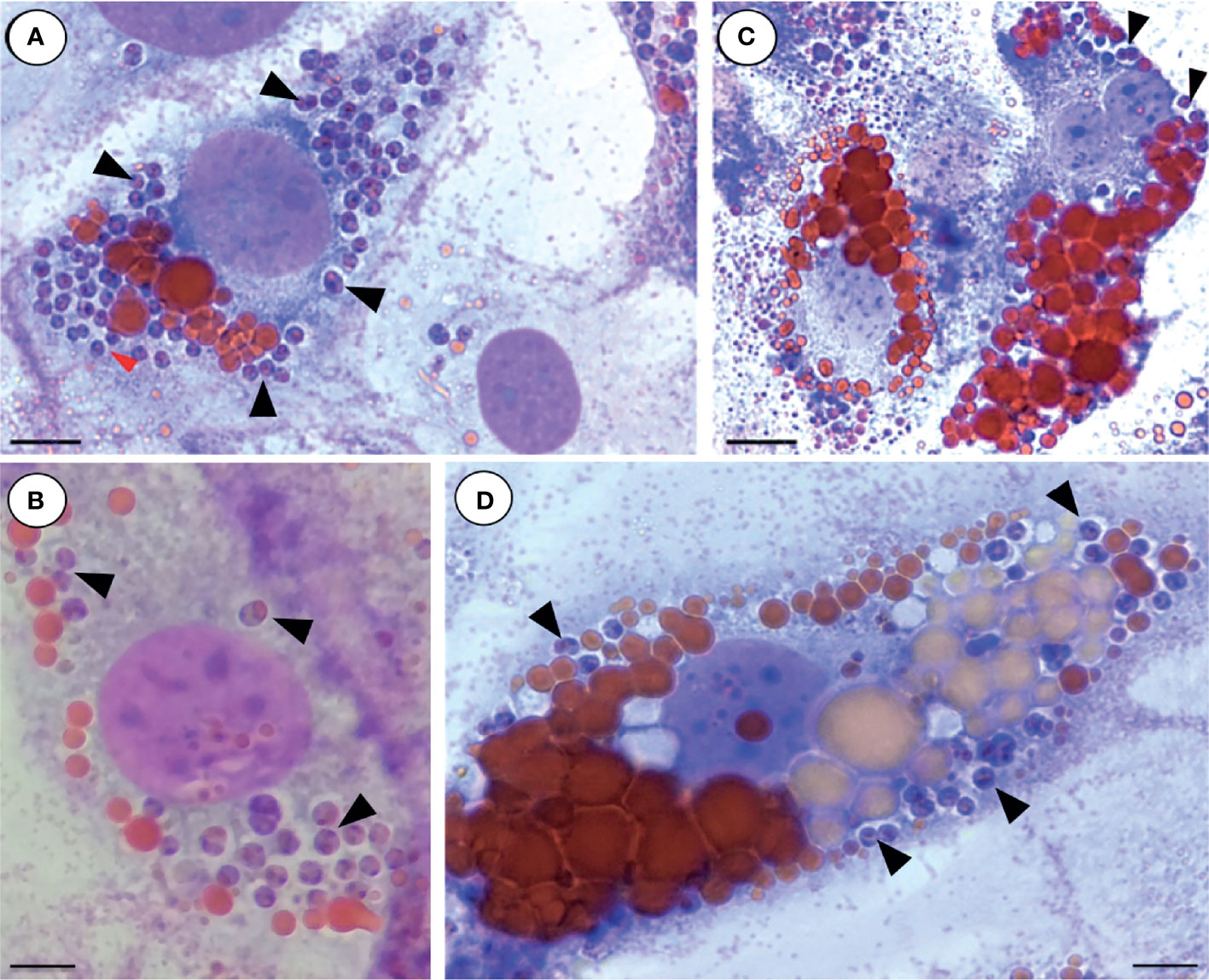
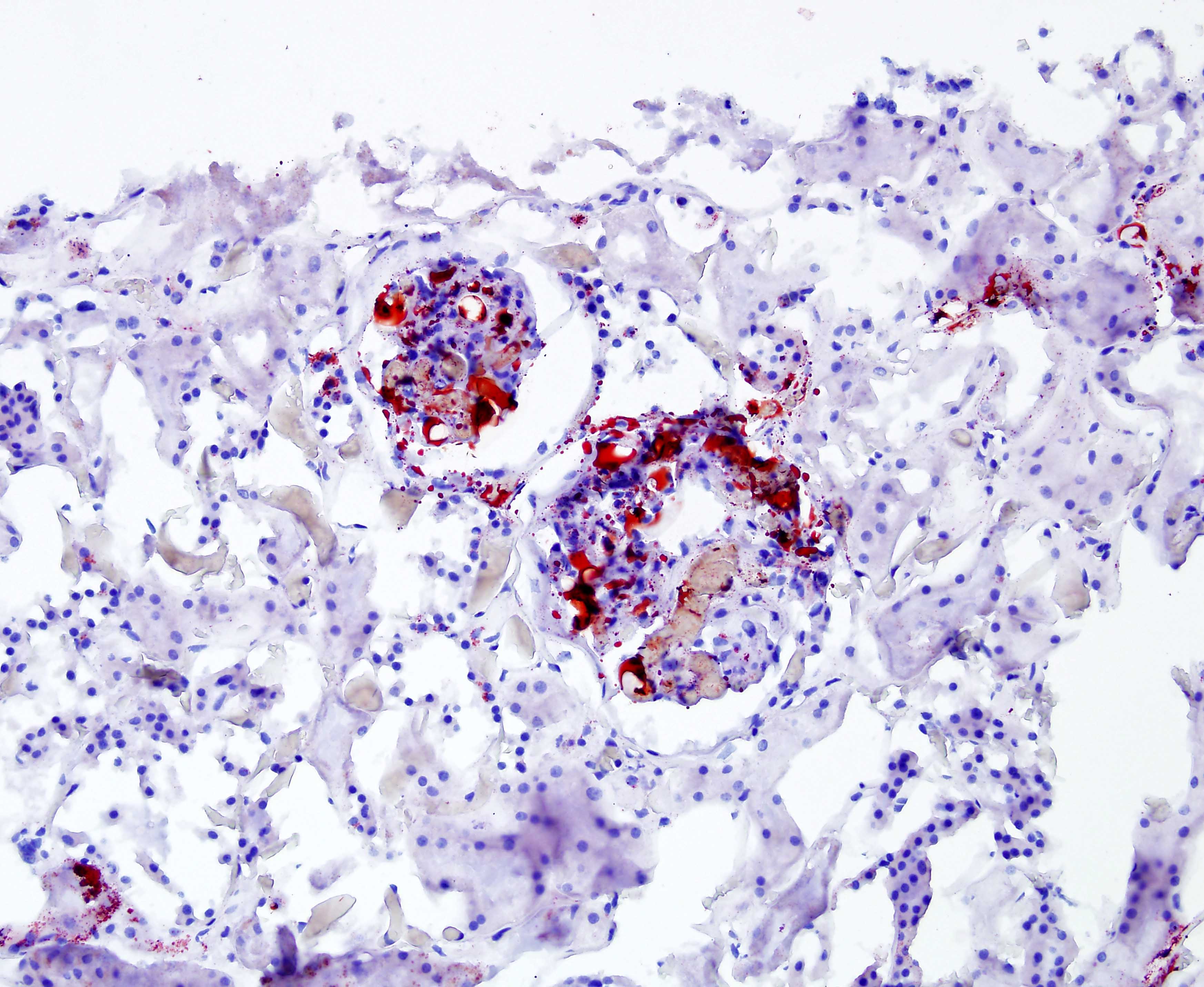
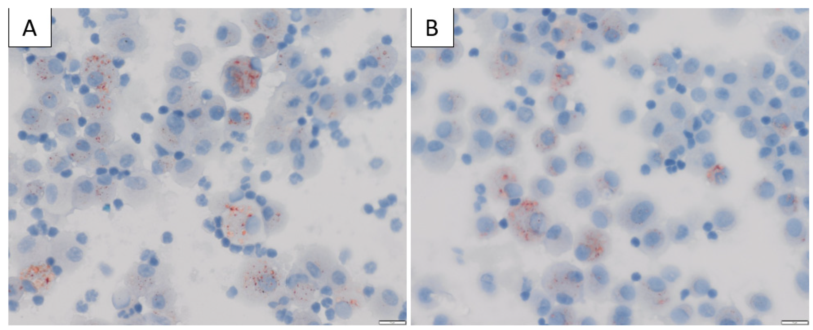
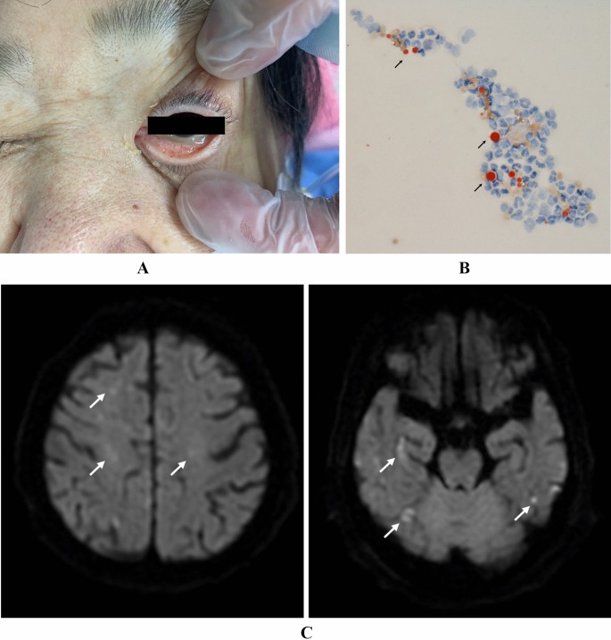
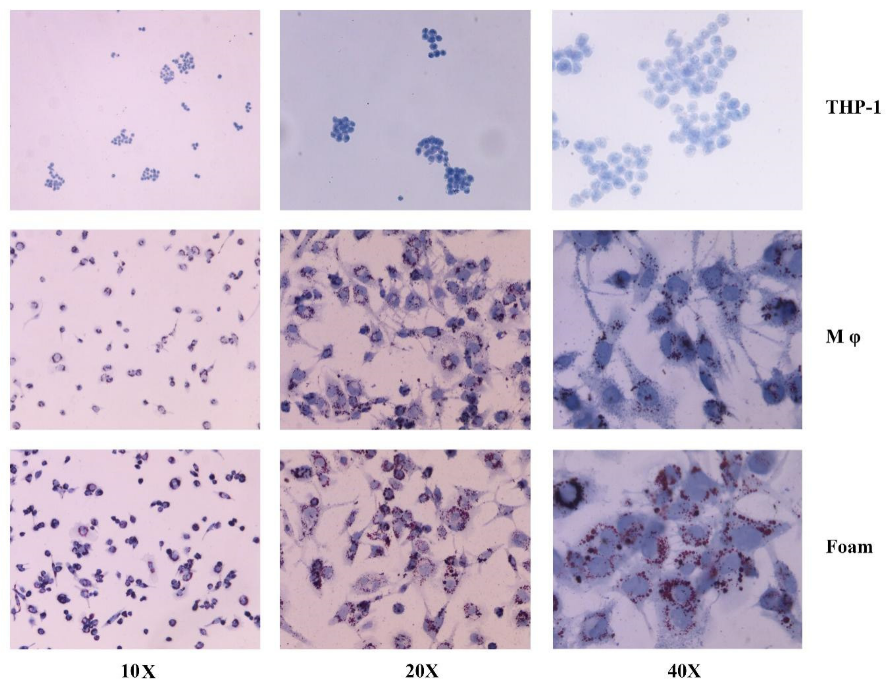
Cytologic specimens from all liposarcomas showed strong positive staining of cytoplasmic vacuoles for lipid Specimens from other sarcomas stained negative for Oil Red O, with the exception of weak, irregular positive staining in 1 hemangiopericytoma Conclusion Our results suggest that Oil Red O staining may be an easy, inexpensive, and useful diagnostic tool for the differentiation of1 Dilute Oil Red O Stock solution 32 using deionized H 2 O to make Oil Red O working solution Example 3mL Oil Red O stock 2mL deionized H 2 O 2 Place a piece of Whatman paper inside the funnel and filter the Oil Red O working solution Alternatively, a syringe filter unit can be used in place of a Whatman paper/funnel system to
Incoming Term: oil red o staining protocol for cytology,
コメント
コメントを投稿